中文

Thondar Product

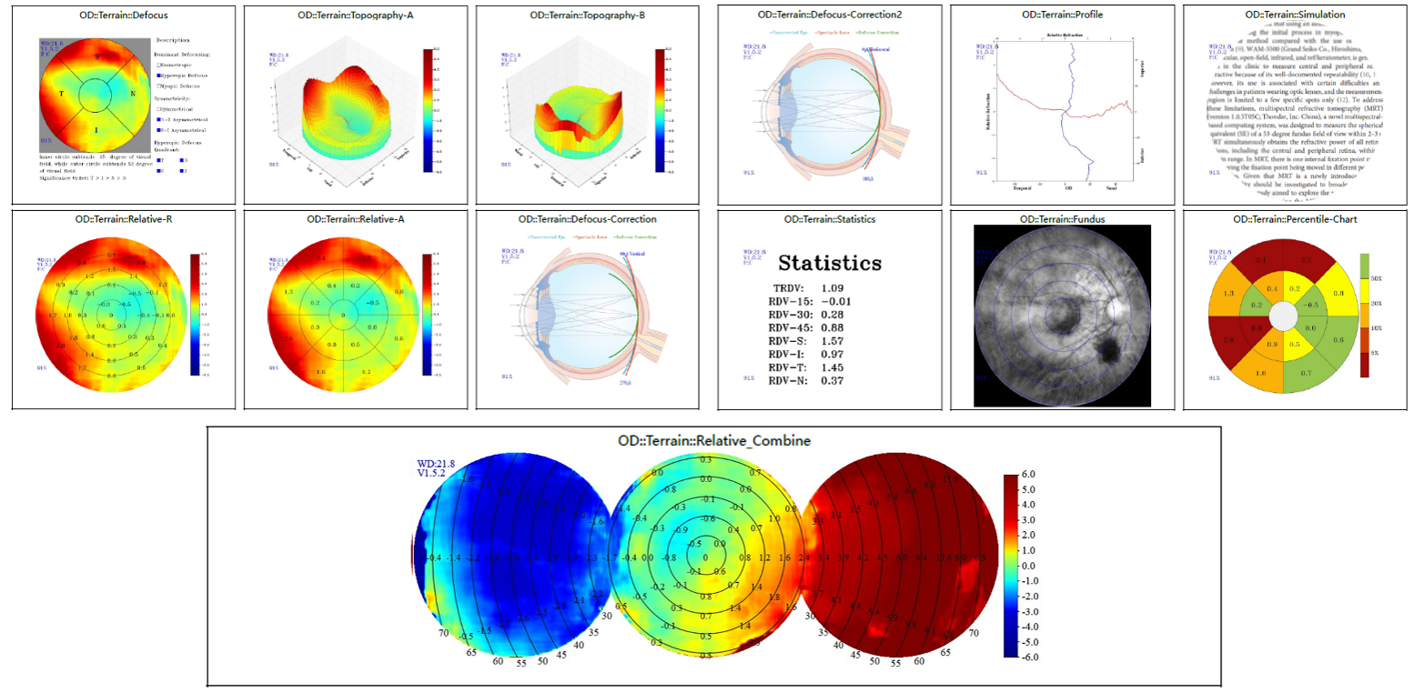
Multispectral Ocular Examination System

Supported by the world's leading technology, it offers multi-model ocular health evaluations and has become a must-have instrument for optometric and ophthalmological practice.


  • Peripheral Refraction Measurement-More Convenient
  • Multispectral Fundus Examination-More Sensitive

PRINCIPLE:

Hyperopic defocus would elongate the axial length of the eye, leading to the  progression of myopia, while an image clearly focused on the retina or in front of it would work as a stop signal for the elongation.


FEATURE:

Continuing the legacy of the Multispectral Refraction Topography ( MRT) technology, it provides RPRE results to evaluate the retinal defocus profile with proven accuracy and repeatability.


SPECIFICATIONS:
Field of View: 53°
Minimum Pupil Size: 3.5mm
Image Pixel: 1000 × 1000
Image Format: JPEG
Power Range: -15D ~ +15D
Illumination Light: LED
Focusing: Auto/Manual
Reports: Defocus, Topography-A, Relative-R, Relative-A, Defocus-Correction, Defocus-Correction2, Simulation, Stastics

Using Multispectral Fundus Imaging(MSI) technology, different levels of fundus image information are collected through a variety of spectra to detect early signs of retinal pathology from shallow to deep, presenting details that are impossible to be captured by ordinary fundus cameras.


Single Spectrum Highlights Lesions,

     Layered Fundus Exploration,
     Deep Choroidal Exploration,
     Multi-dimensional Differential Diagnosis. 


SAMPLE IMAGES:


SPECIFICATIONS:

Output Image: Green to NIR
Illumination Light Spectrum: 488nm ~ 940nm(Peak Wavelength)
Field of View: 63°
Minimum Pupil Size: 3.5mm
Image Pixel: 4096 × 4096
Image Format:JPEG, DICOM
Power Range: -15D ~ +15D
Illumination Light: LED
Focusing: Auto/Manual
Reports: Monochrome Wavelength Image, Color Image